Nanoparticles for Traumatic Brain Injury
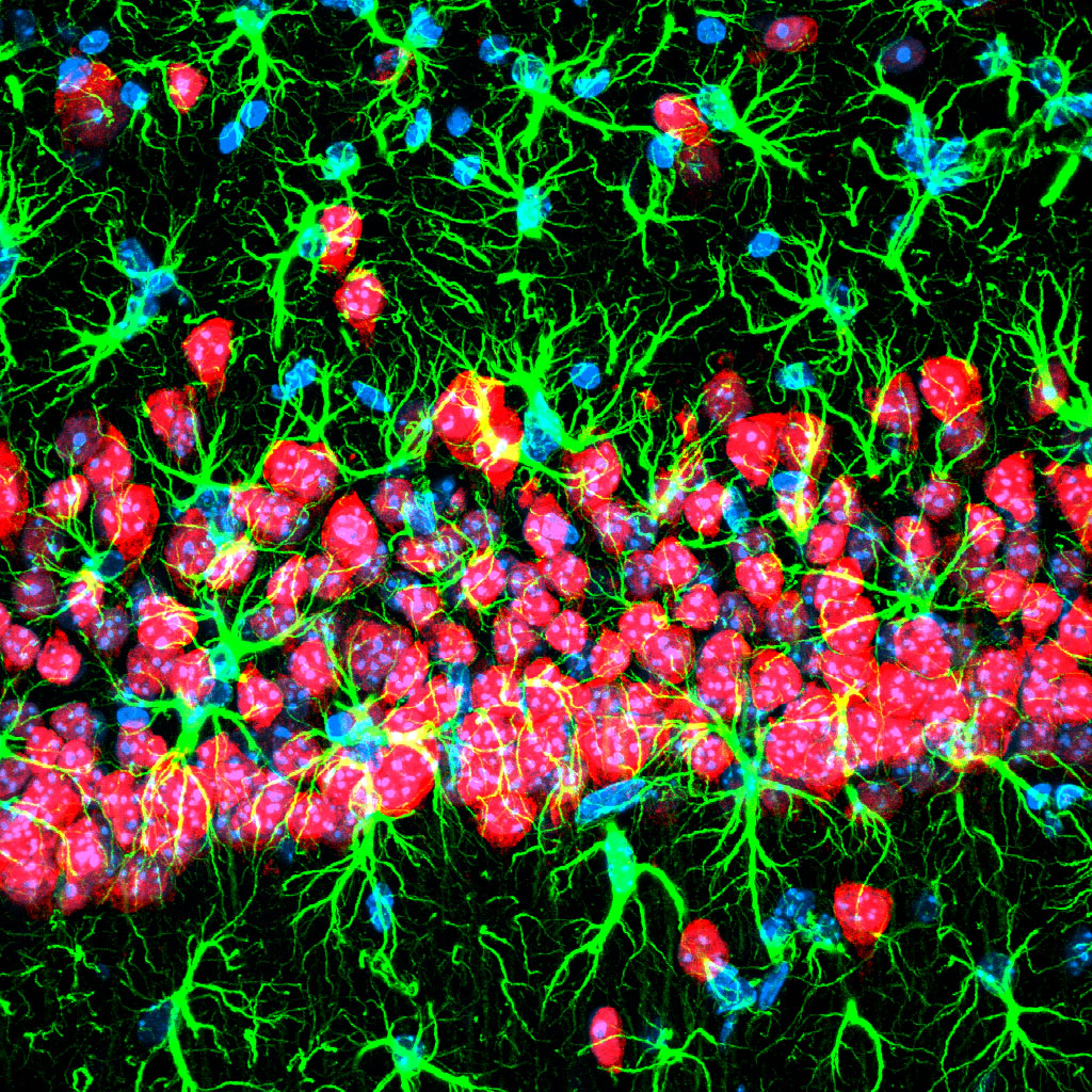
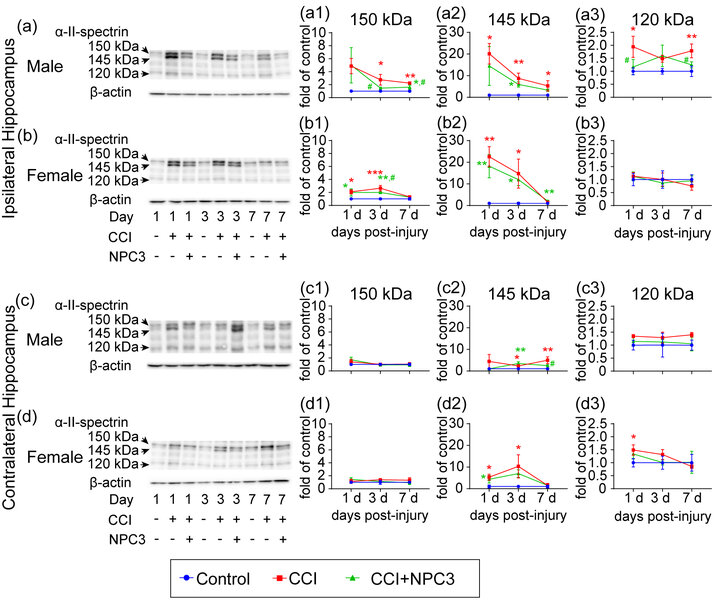
Our nanoparticles for traumatic brain injury (TBI) research goal is to improve treatment of TBI, which has seen little advancement over the past century although it is the leading cause of death and disability in children and adults under 45. We are developing theranostic (combined therapeutic and diagnostic) nanoparticles that can accumulate in damaged brain and reduce long-term secondary effects through histopathological and behavioral analyses in mice.
Nanoparticles for Aging
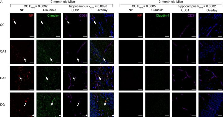
There are numerous diseases related to aging-based neurocognitive decline. Therapy of these diseases is significantly limited by the difficulty in delivery of therapeutics into the brain because of the presence of the blood-brain barrier; thus, a major focus is to improve delivery across this barrier. However, this results in delivery throughout the brain including regions that are normally functioning. We are taking a different approach - our goal is to achieve specific delivery only in regions of brain that are experiencing dysfunction. To do this, we are engineering nanoparticles that can specifically bind to regions of the blood-brain barrier that show endothelial cell surface markers of dysfunction. Our goal improve therapeutic outcomes by using our novel targeted approach to improve therapeutic target engagement.

Nanoparticles for Brain Cancer
We are engineering nanoparticle-based drugs to improve the therapy of pediatric and adult brain cancers. Our goal is to enhance delivery of therapeutics specifically to regions of the brain cancer cells are located and enhancing the effects of radiation and chemo therapies on the cancer cells so that safe doses of therapy that do not harm the healthy brain can be used to kill the tumor.
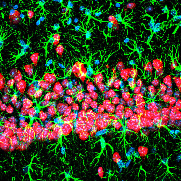
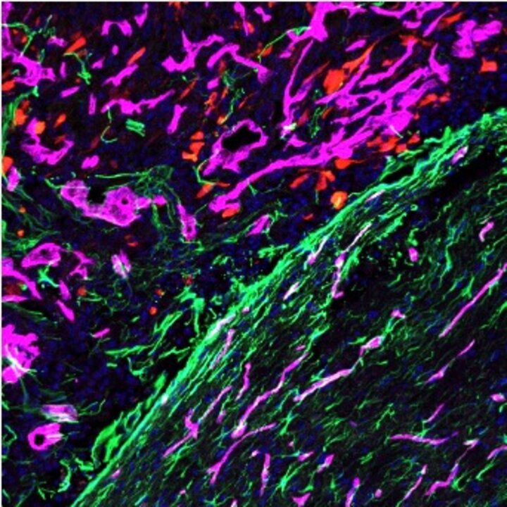
We have found alteration of tight junction protein expression at the blood-brain barrier in response to brain cancer cell signaling and seek to exploit this ectopic tight junction protein expression to improve therapeutic delivery using nanoparticles. Here we see CLDN1 expression (red) in brain tumor tissue but not normal brain. Blood vessels (CD31) are magenta and astrocytes (GFAP) are green.
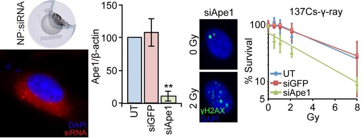
Pediatric brain cancer cells are extraordinarily good at repairing themselves from the damage caused by radiation and chemo therapies. We are developing gene therapy nanoparticles that can inhibit the cell’s ability to repair itself from this damage. These nanoparticles are tiny beads about 1/100,000th the diameter of a human hair. Their small size has allowed us to finely tune their properties at the molecular level to help them deliver treatments specifically to tumors. To the nanoparticles we attach various therapeutics such as siRNA, a small DNA-like molecule, or small molecule drugs that shut down cancer cell repair machinery. These nanoparticles comprise a magnetic contrast agent, which can be imaged using magnetic resonance imaging (MRI) enabling real-time observation of its delivery. This provides a significant advantage over current therapy where delivery cannot be monitored.
Neurobehavioral Testing
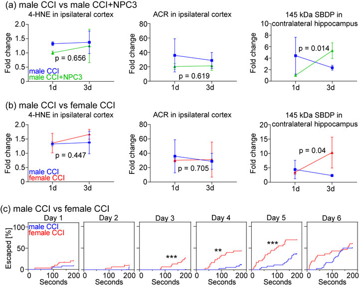
We perform a suite of neurobehavioral testing to test the therapeutic outcome of our NP treatments in our animal models to validate our biomarker findings since changes in biomarker levels does not guarantee improvements in brain function. This includes the Morris water maze, Barnes maze, NSS, elevated plus maze, rotorod, and startle response. We have found significant sex-based differences in spatial learning and memory in response to TBI and NP treatments.


Magnetic Resonance Imaging
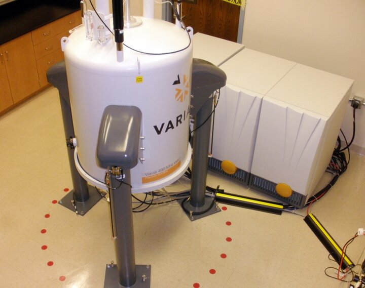
Our lab houses a 9.4T vertical bore Varian magnet for MR imaging of tissues and mice. Please contact us if you're interested in any imaging applications.
High resolution scans of a mouse brain
3D MR image of mouse brain
3D MR image of contrast enhanced brain injury
Diffusion tensor imaging and differential tractography in TBI with NP treatment effects

